The main structures of the eye include:
Cornea: clear tissue in the very front of the eye
Iris: colored part of the eye surrounding the pupil
Pupil: a dark hole in the iris that regulates the amount of light going into the eye
Lens: small clear disk inside the eye that focuses light rays onto the retina
Retina: a layer that lines the back of the eye, senses light, and creates electrical impulses that travel through the optic nerve to the brain.

Macula: a small central area in the retina that allows us to see fine details clearly
Optic nerve: connects the eye to the brain and carries the electrical impulses formed by the retina to the visual cortex of the brain
Vitreous: clear, jelly-like substance that fills the middle of the eye
Glaucoma:
A glaucoma is a group of eye diseases that occur due to elevated intraocular pressure (IOP) inside the eye. The raised pressure affects the optic nerve and may cause vision loss. Glaucoma is classified either as open-angle (more common that is usually painless) or angle-closure glaucoma (which usually occurs suddenly and is associated with pain and redness of the eye).

In the early stages of glaucoma, there are usually no symptoms. By the time vision is affected, the loss is permanent. Progression of glaucoma can be delayed or halted with eye drops, laser treatments, or surgery so early diagnosis is the key.
People with family records of glaucoma, the elderly, and African-Americans are at increased risk of the disease glaucoma.
Cataracts
A cataract is a painless opaque lens in the eye that makes a blurry vision. It progresses gradually as we age. Other causes of cataracts involve diabetes, trauma, some medications, and excessive UV light exposure.

Your physicians can see a cataract during doing a routine eye exam. Treatments for cataracts cover eyeglasses, magnifying lenses, or surgery. Surgery is corrective as the cloudy lens is removed and replaced with an artificial one. The need for surgery and the risks involved should be discussed with your eye physicians.
You Might Also Like:
Constipation :Home Remedies,Symptom,Causes & Medication
Osteoporosis :Symptoms,Cause,Risk factor,Diagnosis,Treatment & Prevention
Gum Disease: Symptom, Causes, Home Remedies & Treatment
Retinal Detachment

Retinal detachment happens when the retina separates from its underlying structures. Retinal detachments are usually painless, and symptoms that may be noticed combine the perception of flashing lights, floaters, or a curtain drawn over the visual field. Risk factors for retinal detachment include adult age 25 to 50, or an elderly person after cataract surgery. Treatment involves surgery, mostly using lasers, that will improve vision affected by the retinal detachment.
Age-Related Macular Degeneration (AMD)
Age-related macular degeneration is an eye disease among onset at any age, normally, after age 60, that progressively damages the macula, the central portion of the retina that aids with focus. It unusually causes total blindness as only the center of vision is affected.

There are two types of AMD: Dry and Wet.
In dry AMD, the light-sensitive cells in the macula slowly break down causing central vision to diminish over time. In wet AMD, abnormal blood vessels behind the retina start to grow, leaking blood and fluid, causing loss of central vision, which may occur quickly.
Conjunctivitis (Pink Eye)
Conjunctivitis is redness and inflammation of the clear tissue covering the eye and the inside of the eyelids known as conjunctiva. It is usually caused by bacterial or viral infections but can also be due to irritants (pollutants, chemicals, or allergens).

Most cases of infections are viral and do not need treatment with antibiotics. Bacterial conjunctivitis can be treated with antibiotic drops or ointments prescribed by the physicians. A crusty discharge can make it difficult to open the eyelids. If this occurs a warm, wet compress may be applied to the eyes to gently remove the crusting.
To decrease the spread of infectious conjunctivitis, wash hands repeatedly, do not share eye drops, cosmetics, towels, or washcloths.
Uveitis
Uveitis is the inflammation of the middle layers of the eye (uvea). The uvea is the layer of the eye which contains the arteries and veins that feed the major structures used in vision. Causes of uveitis involve trauma or injury to the eye, infections, or rheumatologic or inflammatory diseases that attack other parts of the body. The chief symptom of uveitis is the pain in the eyeball. The eye will look red and you will notice blurred vision, light sensitivity, and spots in your vision.

Treatment for uveitis depends on the condition. Anti-inflammatory or antibiotic drops, with pain medications, can be prescribed.
Eye Allergies
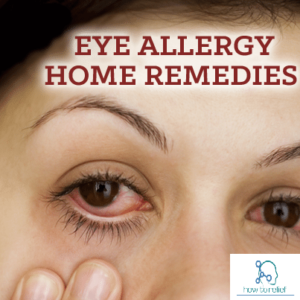
Severe eye allergies can cause damage to the eye that may endanger eyesight. Allergies can induce chronic inflammation that can permanently damage the cornea. Causes of eye allergies are customarily due to seasonal allergies, sensitivities to cosmetics or medications, or dust. Over-the-counter eye drops which contain antihistamines are usually helpful. Consult a physician if OTC remedies do not work, or if you experience pain, discharge, or extreme eye redness.
Sty (Stye)

A sty is an infection of the oil gland at the base of an eyelash. It appears as a red, raised pimple on the edge of the eyelid. Sign & Symptoms of a sty are the pain, redness, tenderness, and swelling with a small pustule. The eyeball itself can feel irritated or as if something is scratching it due to the swelling of the eyelid. Treatment for a sty includes warm compresses applied to the troubled area for 10 minutes, up to six times daily. If the sty comes to a head and releases pus, it should be washed gently with soap and water. This rupture normally leads to the sty going away. If the sty is very painful, large, or affects your vision, see your physicians.
Keratoconus

The cornea is a clear surface coating the front of the eye. It is usually smooth and round, following the contour of the eyeball. Weakness in the structure of the cornea may lead to pressure in the eyeball, causing a conical-shaped irregular bulge to the front of the eye in a condition called keratoconus. Changes in the shape of the cornea do it difficult for the eye to focus even with the help of glasses or contact lenses. Keratoconus may also cause complications during certain eye surgeries. Treatment involves rigid contact lenses or corneal transplantation.
Blepharitis

Blepharitis is an inflammation of the eyelids. The inflammation is found on the inner (posterior) or outer (anterior) eyelid and sign-symptoms include burning, swelling, itching, flaky skin at the base of the lashes, tearing, crusting of the eyelids, or blurred vision. General causes of blepharitis are problems with oil glands at the root of the eyelids, infections, or other skin conditions. Treatment involves good eyelid hygiene, including light scrubbing, frequent cleaning, using a mixture of water and baby shampoo. In the severe cases of blepharitis may require antibiotics or steroids.
Chalazion (Eyelid Cyst)

A
chalazion (also known as a tarsal cyst, meibomian cyst, or conjunctival granuloma) is the inflammation of a small cystic gland in the eyelid. The gland opening becomes clogged and the gland swells. Chalazia are treated with warm compresses, in rare cases, they may require antibiotics. If the chalazion grows severe, causes changes in vision, or is persistent, it may be removed surgically.
Corneal Ulcer

A corneal ulcer is a minute crater (ulcer) on the front part of the eye, customarily resulting from infection. Bacteria, viruses, or fungus may cause a corneal ulcer. People who use contact lenses are at higher risk for corneal ulcers because infectious agents may get trapped abaft a lens. Symptoms of a corneal ulcer include pain, excruciating redness, feeling as if the eye is scratched or something is in the eye, sensitivity to light, and blurry vision. If you doubt a corneal ulcer or have the symptoms of a corneal ulcer and wear contact lenses, optically discern your ophthalmologist immediately. High power antibiotics and pain medications are the treatments for this condition.
Diabetic Retinopathy

People with diabetes often become problems with their blood vessels throughout their bodies and the eye. A complication of diabetes is diabetic retinopathy, that affects the blood vessels in the back of the eye, on the retina.
There are two types of diabetic retinopathy:
Proliferative retinopathy, a severe type where new abnormal blood vessels grow on the retina. These vessels can bleed into the vitreous and cause visual problems.
Nonproliferative retinopathy, a less severe type in that there may be bleeding in the retina and leakage of blood or serum causing a “wet retina.”
Treatment involves laser surgery but the damage will be permanent. The best way to inhibit diabetic retinopathy is with strict glucose control and a healthful lifestyle (dietary restrictions, weight loss, and exercise).
Strabismus (Crossed Eyes)

Crossed eyes or known as strabismus is a condition wherever the eyes do not look in the same direction as they should. One eye may track differently than the other beginning a disjointed appearance. Young children born with this condition may develop decreased vision in one eye (amblyopia). Treatment for strabismus includes using an eye patch on the stronger eye, eye exercises, and possibly surgery.
Floaters

Floaters are induced by aging changes in the vitreous jelly of the eye. They are a normal consequence of aging. If develop multiple floaters or floaters associated with pain, get checked near your ophthalmologist. In general, floaters do not induce blindness and are mostly harmless. There is no definite treatment for floaters, as most will fade or become less noticeable over time.
Farsightedness (Hyperopia)

Farsightedness (hyperopia) is trouble focusing on objects that are close. It is very simple and the incidence increases with age. It is induced by an abnormally flat cornea that does not allow light to sharply focus on the retina. Glasses, contact lenses, or surgery can be used to correct hyperopia.
Nearsightedness (Myopia)

Nearsightedness (myopia) makes people be unable to see distant objects, though people can see nearby objects clearly. It is made by the cornea having too much curvature, resulting in problems with focusing on the retina. Myopia is very common and easily corrected with eyeglasses, contact lenses, or surgery.
Astigmatism

Another usual cause of visual difficulty is astigmatism, in that images are blurred due to an irregularly-shaped cornea. Astigmatism will ultimately affect most people as a part of the aging process. It is managed with glasses, contact lenses, or refractive laser eye surgery.
Color Blindness

The colors we see are a result of how our eyes interpret different wavelengths of light. People including color blindness have difficulty seeing certain colors, usually greens, reds, and blues. Color blindness is generated by an absence or malfunction of color-sensitive cells located in the retina. Maximum of the time this is genetic but it can also be caused by aging, trauma to the eye, disease, or certain medications. If the problem of the color blindness is genetic, the problem cannot be corrected but people can be trained to adapt to interpret color shades. In cases wherever color blindness is acquired, it may be treatable.