The radius is a long bone in the forearm. It lies laterally and parallels to the ulna, It rotates to produce the motion supination and pronation of the forearm. The radius and the ulna to produce movement at the proximal and distal radio-ulnar joints.The radius is part of the elbow and the wrist joints. At the elbow joint, it articulates with the capitulum of the humerus and ulna at the radial notch. At the wrist joint, the radius forms a joint with the ulna bone.
The radius articulates in four areas:
Elbow joint –an articulation(Partly) within the head of the radius, and the capitulum of the humerus.
Proximal radioulnar joint – An articulation of the radial head, and the radial groove of the ulna.
Distal radioulnar joint – An articulation within the ulnar groove and the head of the ulna.
Wrist joint – An articulation among the distal end of the radius and the carpal bones.
Proximal Region of the Radius
- Head of the radius – A disk fashioned structure, with a concave articulating covering. It is thicker medially, wherever it takes part in the proximal radioulnar joint.
- Neck – A small area of bone, which lies within the radial head and radial tuberosity.
- Radial tuberosity – A bony prominence, which serves as the point of attachment of the biceps brachii.
Shaft of the Radius
The radial shaft increases in diameter as it goes distally. Much similar the ulna, it is triangular in shape, with three surfaces and three borders.
In the middle of the lateral surface, there is a small roughening for the attachment for muscle of the pronator teres.
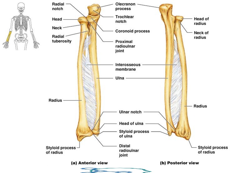
Distal Region of the Radius
In the distal area, the radial shaft extends to form a four-sided end. In the medial surface, there is a concavity, called the ulnar notch, which articulates with the head of the ulna, forming the distal radioulnar joint. The lateral side protrudes distally as the styloid process. The distal surface of the radius has two facets, for articulation with lunate and the scaphoid carpal bones. This forms up the wrist joint.
Muscle Attachments & Movements of the Radius
The biceps muscle inserts on the radial tuberosity of the radius. The upper third of the body of the radius bone attaches to the supinator, the flexor pollicis longus and the flexor digitorum superficialis muscles. The middle third of the body of the radius attaches to the Extensor pollicis brevis muscle, Abductor pollicis longus muscle, and the pronator teres muscles. The lower quarter of the body of the radius attaches to the pronator quadratus muscle and the tendon of the supinator longus.
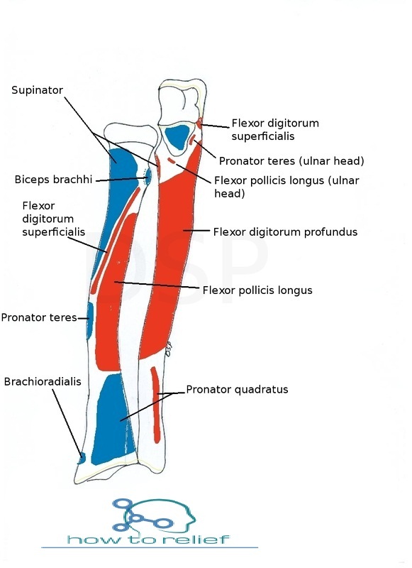
Through supination, the supinator of the forearm and the biceps brachii muscle supinate the forearm by pulling the radius bone. This does the radius move in the opposite direction of the pronator muscles, moving the distal end of the radius back to its point on the lateral side of the wrist.
Through pronation, the distal end of the radius rotates nearby the ulna from its site on the lateral side of the wrist to the medial side of the wrist.
Ossification of the Radius
The radius is ossified from three centers: one for the body and a secondary center for one for either end.
The body of the radius makes its appearance near the center of the bone, through the eighth week of fetal life.
Ossification originates in the lower end between 9 and 26 months of age.The lower epiphysis fuses with the body at the age of twenty.
The ossification center for the upper end seems by the fifth year of age.The lower epiphysis fuses about the age of seventeen or eighteen years.
A further center sometimes seen in the radial tuberosity, develops about the fourteenth or fifteenth year.
Clinical Significance of the Radius
A Proximal radius fracture within the capsule of the elbow joint results in the fat pad sign or “sail sign” which is a displacement of the fat pad at the elbow.
A fracture of the radial head with accompanying dislocation of the distal radio-ulnar joint with separation of the interosseous membrane.
Radial shaft fracture
Distal radius fracture
- A fracture of the radius with dislocation of the distal radioulnar joint, known as Galeazzi fracture.
- A distal fracture of the radius with dorsal displacement of the wrist and hand, known as Colles’ fracture.
- A distal fracture of the radius with volar (ventral) displacement of the wrist and hand, known as Smith’s fracture.
- An intra-articular fracture of the distal radius with dislocation of the radiocarpal joint, known as Barton’s fracture.
- Fracture of radius bone: The radius bone is a weight-bearing bone of the forearm; for this reason fractures of radius bone are more frequent than ulna.
Madelung deformity: It is a congenital defect of radius bone -The anterior bowing of distal end of the radius bone. It occurs within 10 and 14 years of age. There may be subluxation or dislocation of the distal end of ulna, because of defective development of distal radial epiphysis.
Radial aplasia refers to the congenital deficiency or shortness of the radius.