The tibia is the main bone of the leg, It has a proximal and distal end and a shaft, articulating at the knee in proximal and ankle joints in the distal end. It is the second largest bone in the body.
Parts of the Tibia
Proximal End
At the proximal end, the tibia is widened by the medial and lateral condyles. The condyles form a flat surface, known as the tibial plateau.
Medial condyle
Medial condyle is larger than the lateral condyle. Its superior surface connects with the medial condyle of the femur. The articular surface is oval and its large pole is anteroposterior.
The central part of the articular surface is concave and contact with the femoral condyle. The flatter peripheral part separated from the femoral condyle by the medial meniscus. The lateral margin of the articular surface is cover the medial intercondylar tubercle.
On the posterior surface of the medial condyle has a groove. Many vascular foramina mark anterior and medial surfaces.
Lateral Condyle
The lateral condyle is nearly circular [oval in medial condyle] and articulates with lateral condyle of the femur and in peripheral part is covered by the lateral meniscus. The central part is concave just like medial condyle and contact with femoral condyle.
The articular covering has an inflated medial margin which covers the lateral intercondylar tubercle.
The posteroinferior aspect of lateral condyle of the tibia bears a flat, circular fibular facet and is directed downwards, backward and laterally. This facet articulates with the fibula.
The anterior aspect of the condyle bears a flattened impression.
Intercondylar area
The intercondylar area is the roughened area on the superior surface, between the two condyles. This part is elevated to form the intercondylar eminence which is the medial and lateral intercondylar tubercles.
Tibial Tuberosity
Tibial tuberosity is a prominence projection located on the anterior surface of the proximal tibia, inferior to the condyles. In tibial tuberosity where the patella ligament attaches.

Shaft of Tibia
The shaft of the tibia is prismoid in shape and has three surfaces (lateral, medial and posterior) and three borders (anterior, medial and interosseous).
Borders of the shaft of the Tibia
The anterior border is sharp and S-shaped, in the upper part convex medially and convex laterally in the lower part. The anterior border extends from the tibial tuberosity to the anterior border of the medial malleolus. The anterior border forms the shin.
The medial border extends from the medial condyle to the posterior border of the medial malleolus and is rounded.
The lateral border is also described interosseous border and extends from the lateral condyle (just below and in front of the fibular facet) to the anterior border of the fibular notch.
Surfaces of the shaft of the Tibia
The lateral surface rests between the anterior and interosseous borders. In its upper three-fourths it is concave and is directed laterally, and in its lower one-fourth, it is directed forwards.
The medial surface lies within the anterior and interosseous borders. It is subcutaneous in mostly in the upper part.
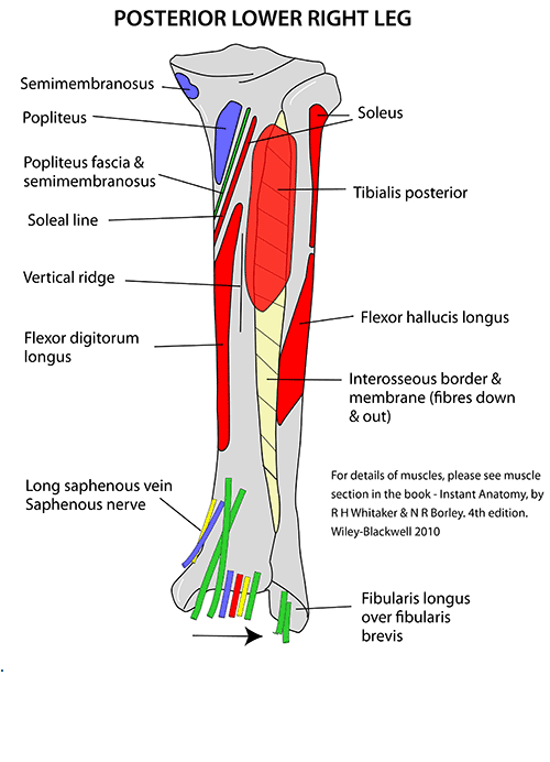
The posterior surface of the tibia is in between medial and interosseous borders. It is widest in its upper part. A rough ridge described as the soleal line crosses it here extending from the fibular facet, running downwards and medially, and terminating by connecting the medial border at the junction of its upper and middle thirds.
Above the soleal line, the posterior surface is in the form of a triangular area whereas the area below the soleal line is elongated and divided into medial and lateral parts by a vertical ridge which regards a downward directed nutrient foramen.
The nutrient foramen transmits the nutrient artery which is a branch of the posterior tibial artery.
Distal End
The distal end of the tibia is expanded but lesser than the upper end. It has a downward projection on the medial side described as a medial malleolus.
It has five surfaces –
The anterior surface of the lower end has an upper smooth part and a lower rough including the grooved part.
The medial surface is subcutaneous and connected with the medial surface of the medial malleolus. The medial malleolus is a little but strong process which projects downwards from the medial surface of the lower end of the tibia.

The lateral surface of the lower end shows a triangular fibular notch to which the lower end of the fibula is attached.
The inferior surface of the lower end of the tibia is articular. It articulates with the superior trochlear surface of the talus and thus takes part in making the ankle joint.
The posterior surface is traversed by a shallow groove directed obliquely downward and medial-ward, assisting for the passage of the tendon of the flexor hallucis longus.
Muscles Attachments of the Tibia
- Tensor fasciae latae muscle insert into the gerdy’s tubercle.
- Quadriceps femoris muscle inserts into the tuberosity of the tibia.
- Sartorius muscle inserts into the pes anserinus.
- Gracilis muscle inserts into the pes anserinus.
- Semitendinosus muscle inserts into the pes anserinus.
- Horizontal head of the semimembranosus muscle inserts into the medial condyle of the tibia.
- Popliteus muscle inserts into the posterior side of the tibia over the soleal line.
- Tibialis anterior muscle arises from the lateral side of the tibia.
- Extensor digitorum longus muscle arises from lateral condyle of the tibia.
- Soleus muscle arises from the posterior side of the tibia under the soleal line.
- Flexor digitorum longus muscle arises from the posterior side of the tibia under the soleal line of the tibia.
Blood supply of the Tibia
The tibia is blood supplied from two sources: as the main source is the nutrient artery, and periosteal vessels arisen from the anterior tibial artery.
Articulation of the Tibia
The tibia is a role of four joints; the ankle, knee, superior and inferior tibiofibular joint.
In the knee joint, the tibia articulates one of the two connections with the femur.This is the weight-bearing part of the knee joint. The tibiofibular joints are the joints of the tibia and fibula which provides very little movement. A proximal tibiofibular joint is a little plane joint. The joint is formed within the undersurface of the lateral tibial condyle and the head of the fibula. The distal tibiofibular joint is formed by the rough, convex surface of the distal end of the medial side of the fibula, and a rough concave covering on the lateral side of the tibia. The ankle joint known as the talocrural joint that connects the distal ends of the tibia and fibula with the proximal end of the talus. The articulation within the tibia and the talus bear more weight than in the smaller fibula and the talus.
Ossification of the Tibia
The tibia is ossified from three centers; a primary center for the shaft of the tibia and a secondary center for one for either end.
- Ossification starts in the center of the body, roughly the seventh week of fetal life, and gradually extends toward the extremities.
- The center for the upper epiphysis appears at close to 34 weeks gestation, lower epiphysis appears in the second year.
- The lower epiphysis fuses with the tibial shaft at approximately the eighteenth, and the upper one fuses approximately the twentieth year.
Clinical Significance of the Tibia
The upper end of the tibia is the most common site of acute osteomyelitis.
- The tibia is normally fractured at the junction of the upper 2/3rd and lower 1/3rd of its shaft.
- The lower two-thirds of the tibial shaft have the low blood supply, the fractures in the lower 1/3rd of the shaft of tibia show delayed union or non-union. Fractures involve the tibia; bumper fracture, Gosselin fracture, Segond fracture, toddler’s fracture, and those including both the tibia and fibula; bimalleolar fracture, trimalleolar fracture, Pott’s fracture.